In our office in Paducah, we utilize a wide variety of technology to diagnose and treat eye diseases. One piece of technology that is often under-utilized by eye doctors is fundus autofluorescence. Here’s a brief explanation of what it is:
The retinal pigment epithelium (RPE) is on the bottom of the 10-layered retina (the light-absorbing tissue that lines the inside of your eye). Within RPE cells, there is a bi-product of retinal metabolism called lipofuscin. Increased or decreased levels of lipofuscin can be markers for specific disease processes.
Very simply put, fundus autofluorescence allows us to take a photograph isolating the RPE. By doing this, it can detect abnormal amounts of lipofuscin.
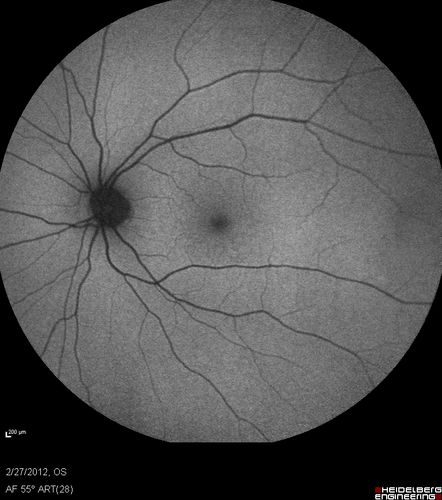
Autofluorescent image of a normal retina
While autofluorescent images may just look like black and white photos, they tell a different story to a trained eye. Excessive amounts of lipofuscin “glow” or “hyperfluoresce” on the images while damaged RPE cells are dark or “hypofluoresce”.
Benefits
Autofluorescent images can pinpoint areas of poorly functioning RPE in certain retinal diseases. These signs often appear on autofluorescent images before they are visible on color photographs. Because of this, autofluorescence is a useful tool to use in addition to color images when monitoring chronic retinal diseases like macular degeneration, histoplasmosis, optic disc drusen, and even malignant melanomas.
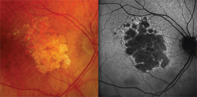
Macular degeneration
We continue to adapt new technology in our practice to better serve our patients in Paducah. By staying current with the latest technology, we can insure that we are providing our patients with the best eye care available.
-Dr. Montgomery
Dr. Canaan Montgomery is an eye doctor in Paducah, KY. As an optometrist, he diagnoses and treats eye diseases, fits contact lenses, and sees pediatric patients.